Anatomy Of Chest X Ray : Chest X-ray / The chest exam is performed more frequently than any other exam in the imaging department.. In fact every radiologst should be an expert in chest film reading. Xray is a type of radiography and most widely used investigation. Roger seheult, md clinical and exam. It is almost always the first imaging study ordered to evaluate for pathologies of the thorax, although further diagnostic imaging, laboratory tests. Each of these anatomical structures should be viewed using a systematic approach.
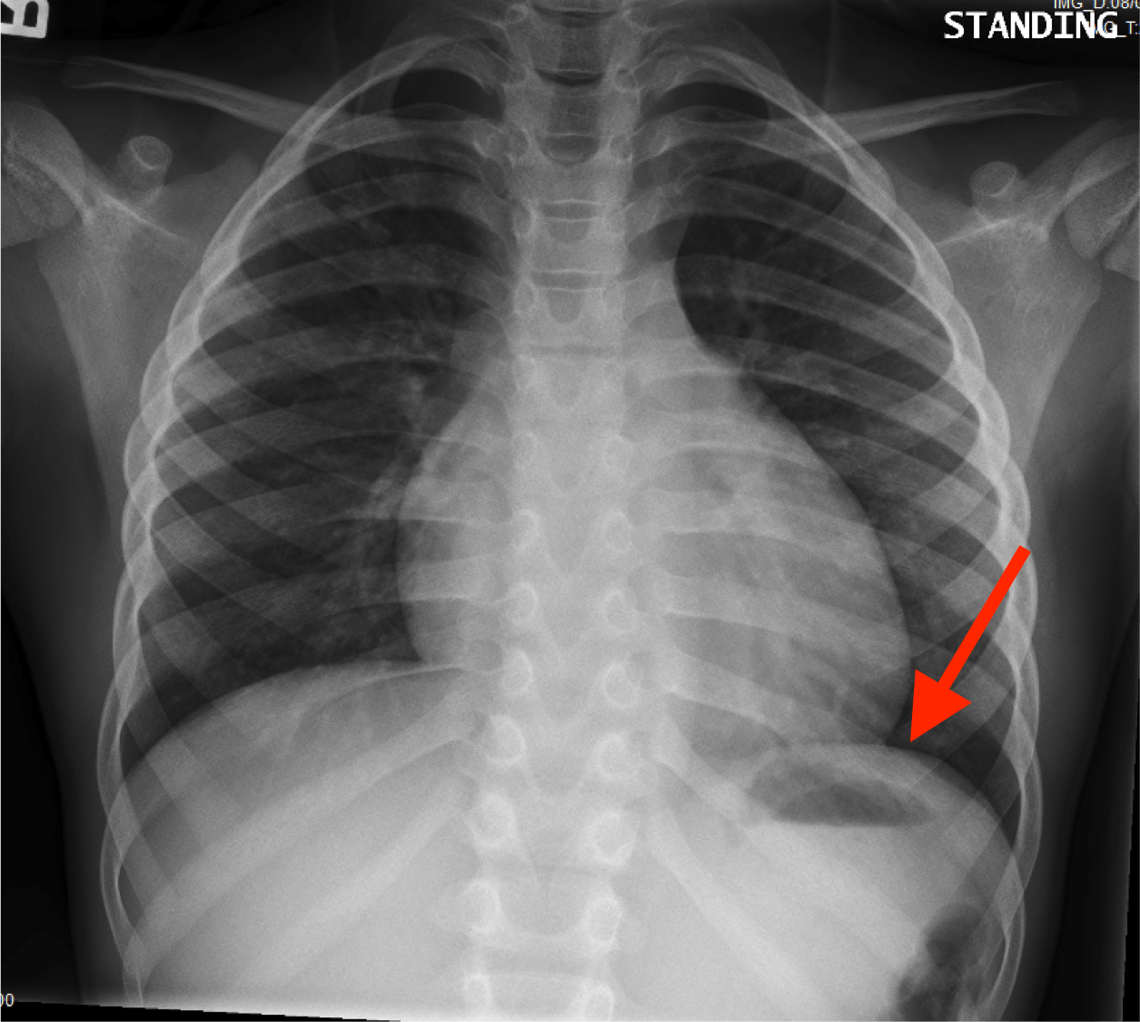
Is there any inhaled foreign body? Submitted 1 year ago by gmdmd. You have completed this module. Major structures are shown in fig. In fact every radiologist and pulmonary physician should be an expert in chest film reading.
Vascular congestion definition & pulmonary vascular congestion from healthjade.net The interpretation of a chest film requires the understanding of basic principles. A collection of anatomy notes covering the key anatomy concepts that medical students need to learn. Note the larger appearing heart on the ap view. Heart abnormalities, including fluid around the heart (pericardial effusion), an enlarged heart (cardiomegaly), heart failure, or abnormal anatomy of the heart can be. In this article we will focus on: Clinicalchest xray anatomy labeled clinical (i.redd.it). Xray is a type of radiography and most widely used investigation. • the straight back syndrome or pectus.
Conclusion of living anatomy of the chest congratulations! Many clinical conditions can be evaluated by this simple radiology test. In fact every radiologist and pulmonary physician should be an expert in chest film reading. Both lungs should be well expanded and similar in volume. In fact every radiologst should be an expert in chest film reading. Chest radiographs are the most common film taken in medicine. Look for lung and pleural pathology. Is there any inhaled foreign body? There are also important structures that are obscured or become visible. Labeled chest radiographs teaching radiologic anatomy with a level of detail appropriate for medical students. Gillian lieberman forthe harvard 62. Doctors use them to diagnose problems. You have completed this module.
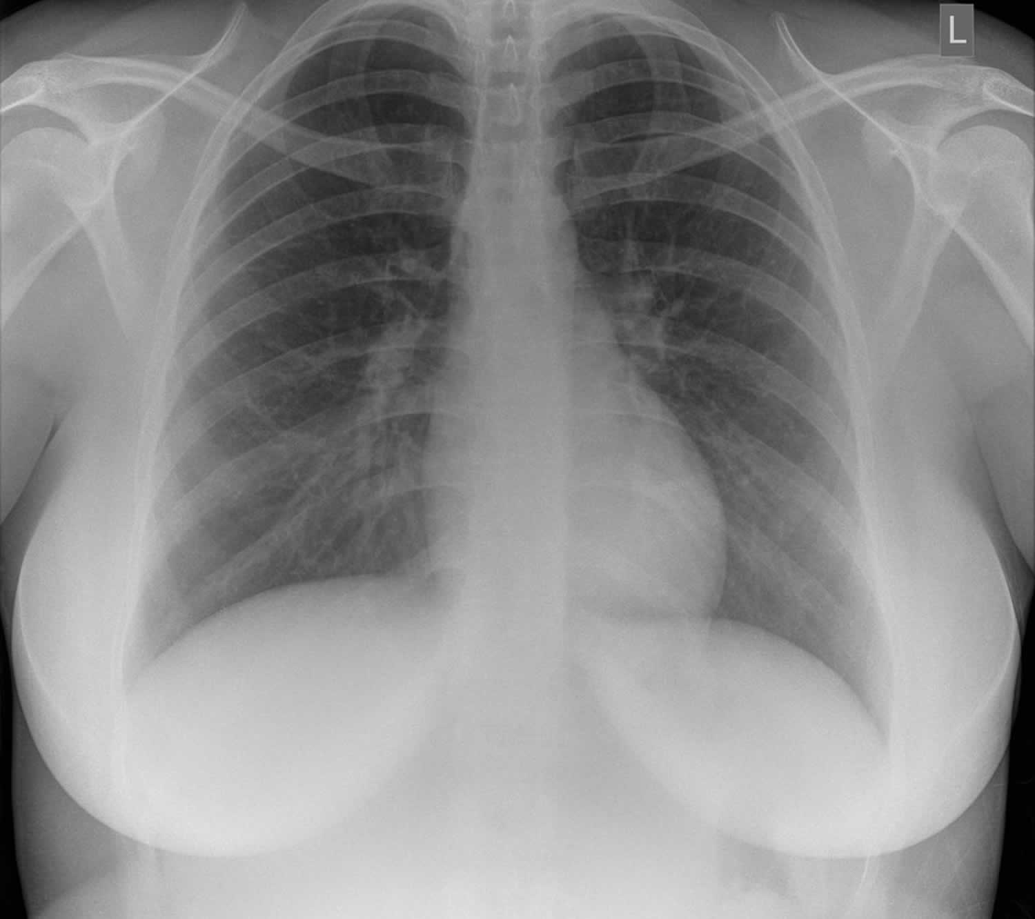
Legit, i can make out the trachea, aorta, outline of the heart, and the diaphragm. There are also important structures that are obscured or become visible. Doctors use them to diagnose problems. The interpretation of a chest film requires the understanding of basic principles. Living anatomy of the chest for 1st year medical students original version compiled by dr.
Normal chest x-ray | Image | Radiopaedia.org from prod-images-static.radiopaedia.org Therefore, knowing the basics and pathologies in the ed setting is very important. A method for examining a chest. Note the larger appearing heart on the ap view. Labeled chest radiographs teaching radiologic anatomy with a level of detail appropriate for medical students. The chest exam is performed more frequently than any other exam in the imaging department. Is there any inhaled foreign body? This imaging method can also check how a patient is responding to specific treatments. Both lungs should be well expanded and similar in volume.
It first appears too complicated to read the chest xrays because we barely know what.
The chest exam is performed more frequently than any other exam in the imaging department. Look for lung and pleural pathology. In fact every radiologist and pulmonary physician should be an expert in chest film reading. Both lungs should be well expanded and similar in volume. This imaging method can also check how a patient is responding to specific treatments. Presence of metallic objects within the area of examination. Abcde aproach comparison of pa vs. Note the larger appearing heart on the ap view. You have completed this module. Gillian lieberman forthe harvard 62. Xray is a type of radiography and most widely used investigation. Roger seheult, md clinical and exam. • the straight back syndrome or pectus.
Structure and function of the shoulder complex. It first appears too complicated to read the chest xrays because we barely know what. A method for examining a chest. There are also important structures that are obscured or become visible. • the straight back syndrome or pectus.
Radiological Anatomy: Stomach - Stepwards from www.stepwards.com This imaging method can also check how a patient is responding to specific treatments. Both lungs should be well expanded and similar in volume. Heart abnormalities, including fluid around the heart (pericardial effusion), an enlarged heart (cardiomegaly), heart failure, or abnormal anatomy of the heart can be. Is there any inhaled foreign body? Many clinical conditions can be evaluated by this simple radiology test. Clinicalchest xray anatomy labeled clinical (i.redd.it). The interpretation of a chest film requires the understanding of basic principles. A free large database of high quality radiology cases with differential diagnoses and mnemonics to help with board.
It is almost always the first imaging study ordered to evaluate for pathologies of the thorax, although further diagnostic imaging, laboratory tests.
The interpretation of a chest film requires the understanding of basic principles. In fact every radiologist and pulmonary physician should be an expert in chest film reading. Living anatomy of the chest for 1st year medical students original version compiled by dr. Heart abnormalities, including fluid around the heart (pericardial effusion), an enlarged heart (cardiomegaly), heart failure, or abnormal anatomy of the heart can be. Xray is a type of radiography and most widely used investigation. In fact every radiologst should be an expert in chest film reading. Therefore, knowing the basics and pathologies in the ed setting is very important. Structure and function of the shoulder complex. In this article we will focus on: It first appears too complicated to read the chest xrays because we barely know what. Submitted 1 year ago by gmdmd. Labeled chest radiographs teaching radiologic anatomy with a level of detail appropriate for medical students. Conclusion of living anatomy of the chest congratulations!
Many clinical conditions can be evaluated by this simple radiology test anatomy of chest. It is almost always the first imaging study ordered to evaluate for pathologies of the thorax, although further diagnostic imaging, laboratory tests.


0 Komentar